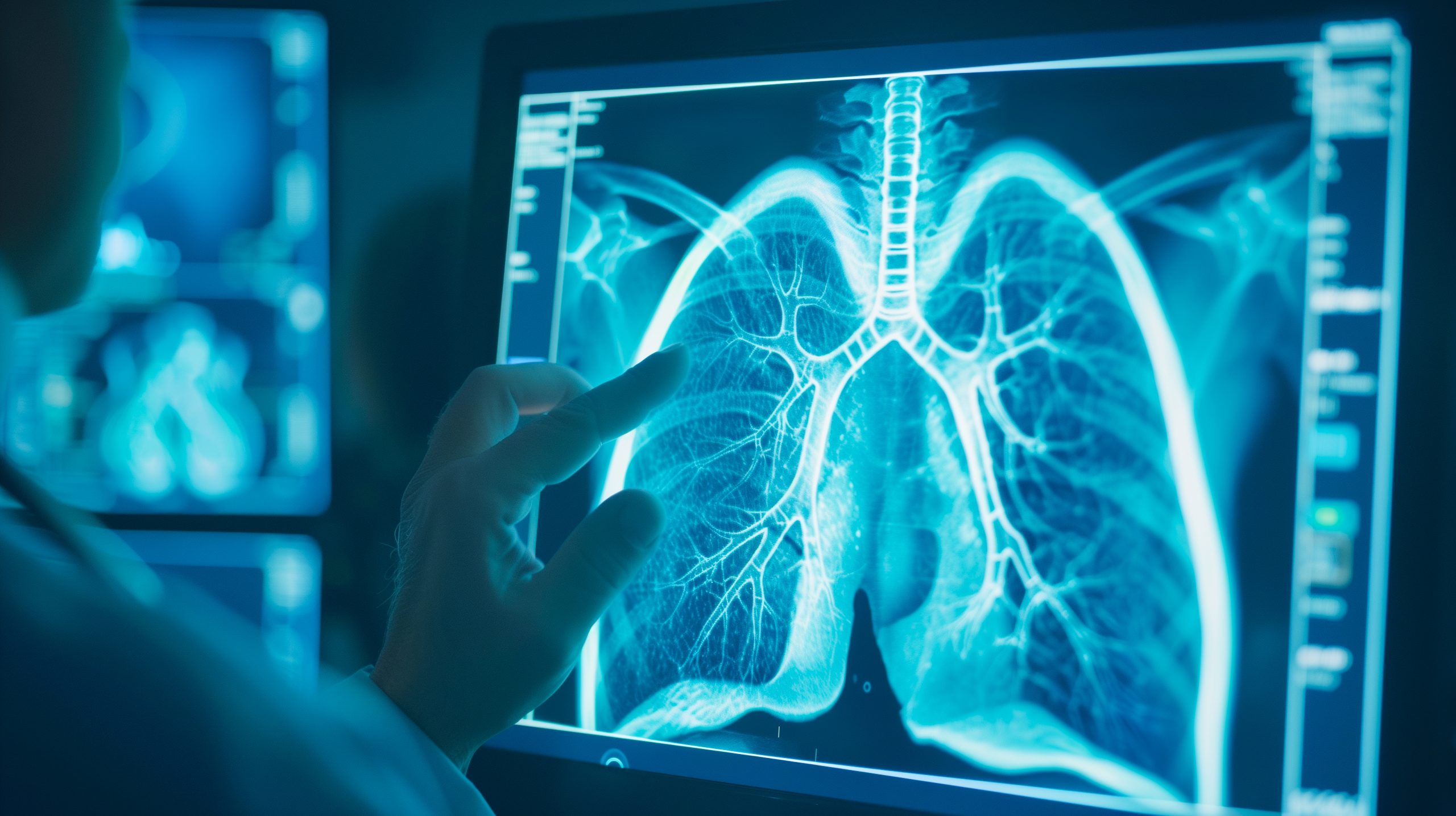
 Chest computed tomography (CT) is essential in the diagnosis of bronchiectasis and offers an invaluable tool to identify patients at risk of poor outcomes.
While bronchiectasis research has advanced and has provided important insights into the epidemiology and pathophysiology of disease, little research has been conducted to understand the radiographic abnormalities found in bronchiectasis patients.
EMBARC is interested in bridging this gap in our understanding and has conducted quantitative analysis of CT scans from bronchiectasis patients enrolled within the EMBARC registry to better understand the nature and extent of structural lung damage/abnormalities in those with bronchiectasis.
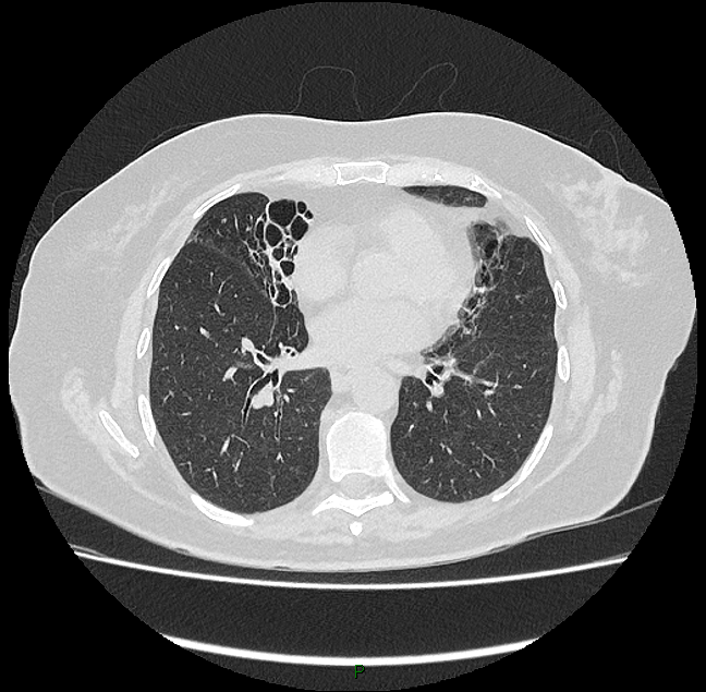
Using an annotated grid-scoring method, EMBARC investigators evaluated CT images from >500 bronchiectasis patients from 8 study sites across 6 European countries. Here, the investigators looked for specific radiologic features including presence and extent of airway widening, consolidation, mucus plugging, airway wall thickening, and parenchymal abnormalities, with a score being calculated to encapsulate the spectrum of radiographic abnormalities.
The study’s findings support the hypothesis that CT imaging can be used to accurately phenotype bronchiectasis and that radiological abnormalities provide valuable clinical insights.
The findings of this study, and an accompanying editorial commentary, are available to read online.